

Beyond the Snapshot: Understanding Longitudinal OCT Imaging

If you've had an OCT scan, you already know how detailed it is. Optical Coherence Tomography (OCT) is a quick, painless imaging test that uses light to create high-resolution cross-sectional images of the retina. It allows your eye care provider to examine the retina and optic nerve in remarkable detail.
A single OCT scan provides valuable information, but it represents only one moment in time. That first scan becomes your baseline. As additional scans are collected over months or years, they are compared with earlier images to determine whether your retina is staying the same or slowly changing over time.
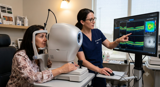
Why One Eye Scan Isn't the Whole Story
Think of a single OCT scan as one chapter in a much longer story. It captures an important moment, but it doesn't reveal how your eye has changed over time.
Comparing two scans provides more information than one, but small differences do not always represent true disease progression. They may simply reflect normal measurement variability, image quality, scan alignment, eye movement, or automated segmentation differences.
In addition, many eye diseases do not progress at a constant rate. Some remain stable for long periods, while others change more rapidly. If follow-up visits are spaced too far apart, important changes may occur between examinations. Every person's eyes are also naturally different. A retinal layer that is thinner or thicker than average is not necessarily a sign of disease. Your previous OCT scans are often the most meaningful reference because they show how your own retina changes over time.
For these reasons, eye care providers look for long-term trends rather than relying on a single measurement.
What Tracking Over Time Reveals
By comparing OCT scans across multiple visits, eye care providers can distinguish normal aging and natural variation from true disease progression. This approach is especially valuable for monitoring chronic eye diseases and evaluating treatment response.
Glaucoma
The retinal nerve fiber layer (RNFL) naturally becomes slightly thinner with age. Serial OCT scans help determine whether the rate of thinning is consistent with normal aging or suggests progressive glaucoma.

Geographic Atrophy
Geographic atrophy, an advanced form of dry age-related macular degeneration, causes progressive loss of the retinal pigment epithelium and photoreceptors. OCT allows these areas to be measured over successive visits, helping determine whether the disease is stable or continuing to progress.
Diabetic Macular Edema
Diabetic macular edema occurs when damaged retinal blood vessels leak fluid into the retina, causing swelling. Because retinal swelling can increase or decrease over weeks or months, repeated OCT scans help assess how well treatment is working and whether additional treatment may be needed.
Evaluating New Treatments
Longitudinal OCT imaging is also an essential tool in clinical research. By measuring structural changes over time, researchers can determine whether a treatment truly slows disease progression rather than simply reflecting normal variation between visits.
Understanding the Limits
Like any medical test, OCT has limitations. Small variations between scans can occasionally suggest a change when none has actually occurred. For that reason, OCT findings are never interpreted in isolation.
If a scan raises concern, your eye care provider will consider your previous OCT scans, your eye examination, your vision, your symptoms, and, when appropriate, other tests before deciding whether a disease is progressing or treatment should change.
In advanced stages of some diseases, OCT measurements may also become less sensitive to further structural change. At that point, other tests, such as visual field testing, may provide additional information about disease progression.
Looking at the Big Picture
A single OCT scan is an important starting point, but the greatest value comes from comparing scans over time.
Longitudinal OCT monitoring helps distinguish normal aging from disease, detect subtle progression before symptoms develop, evaluate how well treatments are working, and support more informed treatment decisions.
Together, these scans provide a clearer and more complete picture of your eye health than any single scan can provide.
This information is intended for general educational purposes and should not replace advice from your own eye care professional regarding your individual eye health or test results.


Comments